- 公司
- 必威体育app官方下载
- 目录
- 新闻和趋势
- 展览
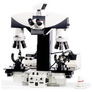
检查显微镜雷声
正直的
3 d
荧光


添加到收藏夹”
这种产品比较
特征
- 技术的应用
- 检查
- 人体工程学
- 正直的
- 观察技术
- 3 d,荧光
- 光源
- LED照明
- 其他特征
- 自动化、图像处理、高速、实时、性价比高
描述
THUNDER Imager Tissue允许实时荧光成像3D组织切片,通常用于神经科学和组织学研究。获得丰富,详细的厚组织图像,从失焦模糊雾霾。即使是组织深处的精细结构也可以通过徕卡创新技术“计算清理”来解决。在大脑切片上成像神经元的轴突和树突等详细的形态学结构。高图像质量,即使是厚的组织切片,与众所周知的速度,荧光效率和易用性的广域显微镜相结合。使用THUNDER成像仪组织为您的研究获得以下优势:•-快速获得无模糊图像,显示最精细的形态学细节,甚至厚部分•深处——让整个组织的快速概述部分•图像和分析具有挑战性的组织与一个简单的工作流细节部分完全解决即使是在厚标本雷声成像仪组织使厚组织标本的意义的探索与所有宽视野显微镜的优点。即使在你的标本深处,也要看到结构的细节。无论你获得的是单个章节还是3D z堆栈。 The investigation of neuronal networks is a perfect example of what is possible. With Computational Clearing, a Leica technique, you remove the out-of-focus blur in real time and make the fine structure of specific brain areas visible. Now you can follow the dynamics of reorganization within a neuronal network or the rebuilding and establishment of new synapses. In other words you can decode 3D biology in real time.
目录
0/10的必威体育app官方下载乘积比较


